New method enables direct observation of bee brains

An international team of bee researchers involving Heinrich Heine University Düsseldorf (HHU) has integrated a calcium sensor into honey bees to enable the study of neural information processing including response to odors. This also provides insights into how social behavior is located in the brain, as the researchers now report in the scientific journal PLOS Biology.
Insects are important so-called model organisms for research. Despite more than 600 million years of independent evolution, insects share more than 60% of their DNA with humans. For several decades it was mainly the fruit fly whose genetic code could be used to study biological processes. Later, such research was expanded to other insects, with particularly promising results coming from the honey bee. Bees display complex social behavior – they perform sophisticated behaviors while employing orientation, communication, learning and memory abilities, which make them interesting subjects for research into the brain's function and neural processing.
A team of researchers from the Universities in Düsseldorf, Frankfurt am Main, Paris-Saclay and Trento has now developed a method to enable direct observation of bee brains, a work which has now been published in PLOS Biology.
A calcium sensor was integrated into the neurons. Calcium plays an important role in nerve cell activity.
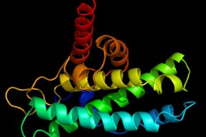
We modified the genetic code of honey bees to make their brain cells produce a fluorescent protein, a sort of sensor that allows us to monitor the areas that are activated in response to environmental stimuli. The intensity of the light emitted varies according to neural activity."
Dr Albrecht Haase, Professor of Neurophysics, University of Trento
Omics eBook

Professor Beye indicates that "the realization of this "sensor bee" was particularly challenging because we had to work on the DNA of queen bees. Unlike fruit flies, the queen bee cannot easily be maintained in the laboratory, because each one needs its own colony."
The research started with the inoculation of a specific genetic sequence into over 4,000 bee eggs. The protracted breeding, testing and selection process ultimately resulted in seven queens carrying the genetic sensor. When they reproduced in their own colony, the queens transmitted the gene to some of their offspring.
The sensor developed by the team of researchers was then used to study the bees' sense of smell and how the perception of smell is encoded in the neurons. Dr Julie Carcaud, Assistant Professor at the University of Paris-Saclay and Dr Jean-Christophe Sandoz, Research Director at CNRS in Paris, explain: "The insects were stimulated with various odors and observed with a high-resolution microscope. This made it possible to detect which brain cells are activated by these smells and how this information is distributed in the brain."
Dr Marianne Otte, co-author of the study from Düsseldorf: "The recordings were performed in vivo using techniques which enabled us to look into the brains of the bees. The insects were fixed in a measuring stand and then presented with various odor stimuli."
Professor Dr Bernd Grünewald from Goethe University Frankfurt am Main and Director of the Honeybee Research Center in Oberursel: "The new "sensor bee" makes it possible to study how communication works within colonies and, more generally, how sociality affects the animals' brains."
Heinrich-Heine University Duesseldorf
Carcaud, J., et al. (2023) Multisite imaging of neural activity using a genetically encoded calcium sensor in the honey bee. PLOS Biology. doi.org/10.1371/journal.pbio.3001984.
Posted in: Molecular & Structural Biology | Histology & Microscopy
Tags: Bee, Brain, Calcium, Cell, DNA, Evolution, Fluorescent Protein, Fruit, Gene, Genetic, Honey, Imaging, in vivo, Laboratory, Microscope, Model Organisms, Nerve, Neurons, Protein, Research
Source: Read Full Article