Raman Imaging Techniques

Raman imaging techniques are used to obtain information about the molecular vibrations and crystal structure of a sample. Image pixels are used to acquire a complete spectrum, which is subsequently interrogated in order to generate color images that differ according to material, crystallinity, phase, and strain. Several variations of raman imaging exist, and these are explored in this article.
Skip to:
- CARS (Coherent anti-stokes Raman spectroscopy)
- SERS (Surface Enhanced Raman spectroscopy)
- Raman spectroscopy with Single-Walled Carbon Nanotubes (SWNTs)
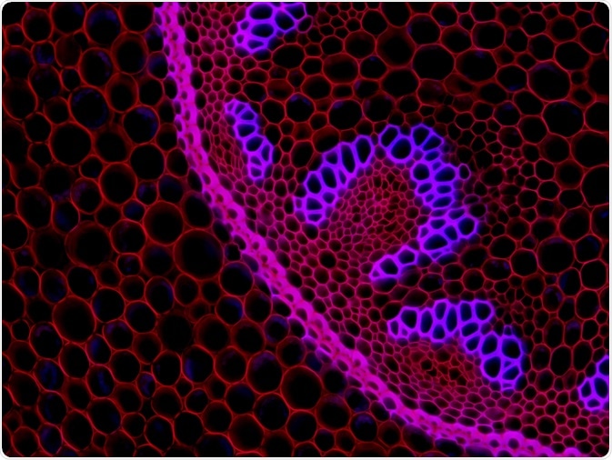 Virginie Thomas | Shutterstock
Raman spectroscopy
In Raman spectroscopy, a laser is used to irradiate a sample. Almost all of the light is scattered in an elastic manner, meaning no change in energy occurs. However, a small percentage of light is scattered in an inelastic manner, known as Raman scattering. This process was first observed by Indian scientist Sir Chandrashekhara Venkata Raman in 1928.
When the incident light interacts with a molecule, the electron absorbs that energy and moves to an excited state. Still, this state is not stable and it comes back to the original state with the same previous energy levels. This is called Rayleigh scattering (whereas, in Raman scattering, the energy levels are different compared to the original state).
In Stokes scattering, the electron gets excited and then comes to a vibrational level by absorption of energy. Thus, the molecule has lower energy and longer wavelength compared to the original incident light.
On the other hand, in anti-Stokes scattering the electron comes from the vibrational state to the ground state, and there is an energy transfer to the scattered photon. Thus, the Raman scattered light has more energy and shorter wavelengths compared to the incident light.
CARS (Coherent anti-stokes Raman spectroscopy)
CARS is a form of Raman spectroscopy that is used to detect the vibrational signatures of molecules – including the vibrations of chemical bonds. This method uses multiple photons to detect the molecular vibrations, producing a signal where the emitted rays are coherent to each other. This signal is enhanced when the difference in the frequencies of the two laser pulses is equal to the Raman frequency of the chemical bond that is under study.
CARS is mainly used to detect molecules in biological tissues and lipids that have a high density of CH2 groups. Inside cells, this is used to image intracellular transport of organelles, intracellular lipid molecules, as well as to differentiate stem cells from other cells in the culture. In one study CARS was combined with video-rate microscopy which led to real-time imaging of dynamic processes, including diffusion of chemical molecules in tissues.
SERS (Surface Enhanced Raman spectroscopy)
SERS is a method that enhances the Raman scattering using adsorbed molecules on the surface or nanostructures, including plasmonic-silica nanotubes up to a factor of 1010. This method can be used to detect single molecules, and the adsorbed molecules can increase the Raman scattering by increasing the incident electric field.
Usually, metal nanoparticles, such as gold and fluorescent dyes, are used for SERS-based imaging. In vivo imaging has also been performed with SERS imaging in animal models. For example, SERS nanoparticles were conjugated with tumor-targeting ligands for imaging purposes.
Raman spectroscopy with Single-Walled Carbon Nanotubes (SWNTs)
Single-walled carbon nanotubes (SWNTs) have a strong Raman peak that is produced by the coupling of electron and photons. This leads to excitation of vibration when the light is incident. The Raman peak of SWNTs are narrow and easily identified from the background of autofluorescence.
Unlike the SERS method, this technique does not require the presence of metal nanoparticles to enhances its detection. This method has also been used to perform Raman imaging of live cells. In another study, antibodies functionalized with SWNTs were used as multicolor Raman label for sensitive and multiplexed detection of proteins.
Sources
- www.ncbi.nlm.nih.gov/pmc/articles/PMC2917525/pdf/nihms-186152.pdf
- web.pdx.edu/…/Raman_Spectroscopy_Basics_PRINCETON-INSTRUMENTS.pdf
- www.physik.fu-berlin.de/einrichtungen/ag/ag-reich/docs/Thomsen2007.pdf
- https://www.mdpi.com/2079-6374/9/1/25
Further Reading
- All Raman Spectroscopy Content
- Raman Spectroscopy: Factors that Affect Spectral Resolution
- Transmission Raman Spectroscopy Advantages and Limitations
- What is Confocal Raman Microscopy?
- Using a Portable Raman Spectrometer for Detection of THC
Last Updated: May 9, 2019

Written by
Dr. Surat P
Dr. Surat graduated with a Ph.D. in Cell Biology and Mechanobiology from the Tata Institute of Fundamental Research (Mumbai, India) in 2016. Prior to her Ph.D., Surat studied for a Bachelor of Science (B.Sc.) degree in Zoology, during which she was the recipient of anIndian Academy of SciencesSummer Fellowship to study the proteins involved in AIDs. She produces feature articles on a wide range of topics, such as medical ethics, data manipulation, pseudoscience and superstition, education, and human evolution. She is passionate about science communication and writes articles covering all areas of the life sciences.
Source: Read Full Article