How a single synapse transmits both visual and subconscious information to the brain of fruit flies


Research led by Peking University, China, has discovered a single type of retinal photoreceptor cell in Drosophila (fruit fly) is involved in both visual perception and circadian photoentrainment by co-releasing histamine and acetylcholine at the first visual synapse.
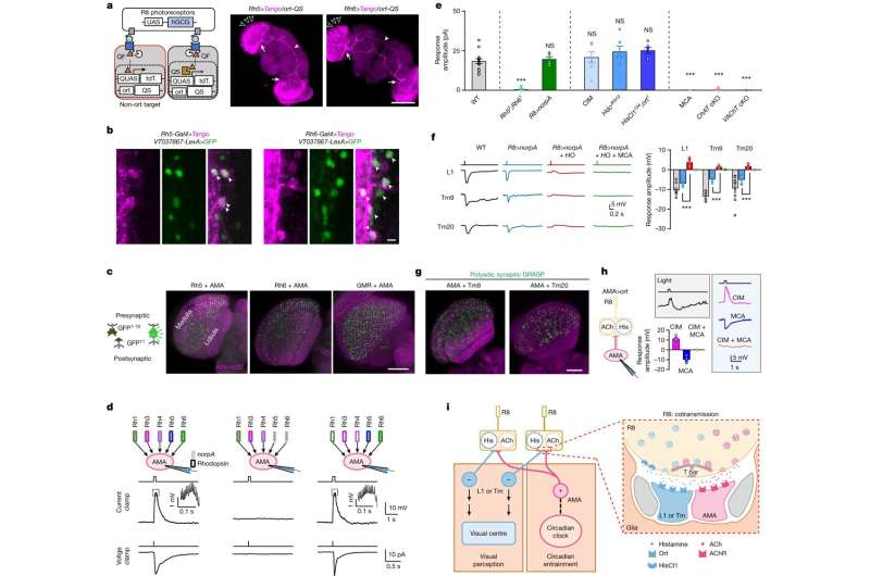
In a paper, “A single photoreceptor splits perception and entrainment by cotransmission,” published in Nature, the team details the discovery that the Drosophila visual system segregates visual perception and circadian photoentrainment by co-transmitting two neurotransmitters, histamine and acetylcholine, in the R8 photoreceptor cells.
Light detection involves capturing signals through photoreceptors in the eye, which are essential for image formation and subconscious visual functions, such as regulating biological rhythms according to the daily light-dark cycle (photoentrainment of the circadian clock). The optical system has distinct pathways for image formation (based on local contrast) and non-image-related tasks (based on global irradiance).
The study uncovers a neural basis for the separation of visual signals. It demonstrates that image-related and subconscious sensory functions can be separated at the first synapse of a system that transmits two chemical messengers (histamine and acetylcholine).
The R8 photoreceptors in Drosophila were found to be responsible for both image-forming vision and circadian photoentrainment. When light is detected, R8 co-transmits two different neurotransmitters, histamine and acetylcholine, setting off different functions. A contrast-based pathway drove the image-forming vision pathway, while the circadian photoentrainment relied on an irradiance-based pathway.
Fruit flies (Drosophila melanogaster) are often used as a model for vision research. The study involved measuring ion currents in individual cells in the fruit fly brain responsible for regulating the circadian clock. Only specific photoreceptors, the blue-sensitive pR8 and green-sensitive yR8, were found to transmit irradiance signals to the central clock neurons in the fruit fly brain using the neurotransmitter acetylcholine.
Eliminating histamine and acetylcholine transmission impaired motion detection and circadian photoentrainment, respectively. This demonstrates the importance of these neurotransmitters in driving specific behaviors.
Specific clock neurons (labeled AMA for this study) receive irradiance inputs from both pR8 and yR8 photoreceptors. These AMA neurons integrate irradiance inputs from different directions and wavelengths of light and are connected both chemically and electrically.
The authors suggest that conventional photoreceptors other than R8 may also use histamine-mediated pathways to excite clock neurons for circadian photoentrainment. These pathways might reconstruct irradiance signals from image-forming signals, an unexpected crosstalk between image-forming vision and circadian photoentrainment, highlighting the dynamic regulation of neurotransmitter release in response to light stimulation.
While this study sheds light on the mechanisms that allow the fruit fly to distinguish between different visual functions, it suggests that similar mechanisms could be at work in the visual pathways of mammals. Identifying the circuits that extract irradiance signals from image-forming pathways could enhance our understanding of visual processing. The study also offers insights into how dim light can activate clock neurons even without histamine signaling.
More information:
Na Xiao et al, A single photoreceptor splits perception and entrainment by cotransmission, Nature (2023). DOI: 10.1038/s41586-023-06681-6
How light receptor cells in fruit-fly eyes multitask, Nature (2023). DOI: 10.1038/d41586-023-03268-z
Journal information:
Nature
Source: Read Full Article