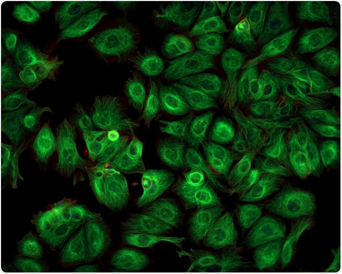
Total Internal Reflection Fluorescence (TIRF) Microscopy Applications

Total internal reflection fluorescence (usually abbreviated as TIRF) microscopy is an imaging method that illuminates fluorescence in a shallow area of the specimen in order to enhance the signal-to-noise ratio. This technique is widely applied in imaging small structures or molecules.

Caleb Foster | Shutterstock
Methodology of TIRF microscopy
In TIRF microscopy, fluorescent molecules are in a sample in an aqueous environment that is near a solid with a high refractive index, usually a glass coverslip. At what is called the critical angle, the illumination is totally reflected off the interface between the glass and the liquid. This reflection creates a thin electromagnetic field called the evanescent wave.
The evanescent wave excited fluorophores within 100-200 nm of the glass coverslip. This thin field is important for three reasons: the signal-to-background ratio is high, very little fluorescence that is out of focus is collected, and lastly, the light that cells are exposed to is substantially less than traditional microscopy techniques.
Basic applications of TIRF microscopy
TIRF microscopy was initially developed in the 1980s, but has not been used much until recently. In the early days, TIRF was used to investigate the contact areas of cells and substrates of human skin fibroblasts, along with studies on biomedical surface dynamics and energy transfer in cells.
The main uses of TIRF microscopy are related to the cell membrane, since this is the area that is primarily imaged. Consequently, TIRF microscopy can be employed to record the behavior of ligand-mediated receptor endocytosis, as well as the lateral movement of receptors along the membrane. The receptors in such cases are usually imaged using fluorescently tagged ligands, antibodies, or small molecules.
TIRF microscopy has also been applied to study exocytosis and endocytosis. Fluorescent dyes or proteins can be embedded with the cargo of a vesicle, making it possible to track the vesicle’s movement during exocytosis.
TIRF microscopy has similarly been applied to endocytosis, where it has helped in defining the roles of proteins such as Rab3A and Rab27A, showing that those two proteins regulate docking steps of the vesicle to the membrane in a cooperative manner. It was also shown how Rab3A will dissociate from a vesicle during exocytosis.
The excitation of fluorophores via the evanescent field is highly useful for thicker cells, such as mammalian cells. Mammalian cells also contain naturally occurring fluorophores, such as flavin and NADH. The thin section achieved by TIRF microscopy ensures that such naturally occurring fluorophores are not significantly excited. This helps elevate the high signal-to-noise ratio.
Optical sectioning of this kind makes TIRF microscopy ideal for imaging molecules and features at the interface between the cell surface and substrate, for example focal adhesions, secretory vesicles, as well as early endocytic particles.
Can TIRF be applied to neuroscience?
TIRF microscopy is limited to imaging structures and processes happening at or close to the interface between the specimen and the coverslip. However, there are several important applications where questions near the cell surface need thorough investigation, such as neuroscience questions.
The release and uptake of neurotransmitters at neuronal synapses have previously been studied by ways that can be argued to be indirect, such as genetic and electron microscopic approaches.
On the other hand, TIRF microscopy offers direct visual imaging of dynamic processes such as the protein and vesicle interactions in neurons. For examples, studies have visualized the release of vesicles containing lipids from active zones, and the transport of vesicles from a reserve pool 20 nanometers away from the plasma membrane to replace the released ones.
Complex uses and considerations
In 2011, further developments of this method led to better imaging of features deeper inside the cell. The total internal reflection which generates the evanescent wave usually occurs at the interface between the sample and the glass coverslip, but the interface can be located deeper in the sample.
The basis of generating the evanescent wave is that two surfaces (sample and glass) need to have different refractive indices. If the interface between media with different refractive indices occurs within the sample, the imaging can be done deeper in the cell. For example, the plasma membrane in plant cells has been amenable to such imaging.
Another a similar concept can occur that is called ‘frustrated TIRF’. In it, the field that is created at the interface between the coverslip and the sample propagates light to a second interface. This interface is located within the evanescent field.
The frustrated TIRF can be created if the refractive indices in a sample are at a certain value. Thankfully, frustrated TIRF can be told apart from normal TIRF by that the beam reflected from the interface is significantly weaker.
Sources
- https://www.ncbi.nlm.nih.gov/pmc/articles/PMC4540339/
- www.microscopyu.com/…/total-internal-reflection-fluorescence-tirf-microscopy
- https://www.ncbi.nlm.nih.gov/pmc/articles/PMC4285862/
Further Reading
- All TIRF Microscopy Content
- TIRF Microscopy Explained
Last Updated: Nov 27, 2018

Written by
Sara Ryding
Sara is a passionate life sciences writer who specializes in zoology and ornithology. She is currently completing a Ph.D. at Deakin University in Australia which focuses on how the beaks of birds change with global warming.
Source: Read Full Article