A better map of the lights you see when you close your eyes can improve bionic eye outcomes


Researchers at Monash University have identified a new way of mapping ‘phosphenes’—the visual perception of the bright flashes we see when no light is entering the eye—to improve the outcome of surgery for patients receiving a cortical visual prosthesis (‘bionic eye’).
Cortical visual prostheses are devices implanted onto the brain with the aim of restoring sight by directly stimulating the area responsible for vision, the visual cortex, bypassing damage to the retina of the eye or the optic nerve.
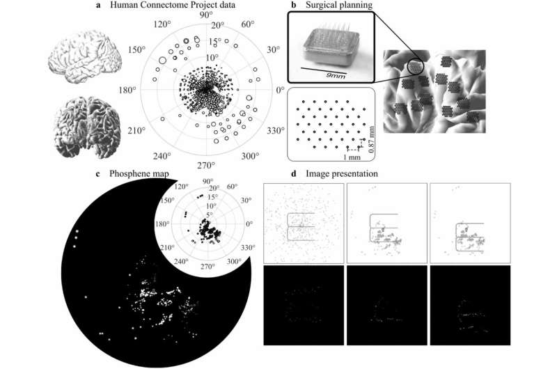
A typical prosthesis consists of an array of fine electrodes, each of which is designed to trigger a phosphene. Given the limited number of electrodes, understanding how electrodes can best be placed to generate useful perceived images becomes critical.
As part of this researchers from the Department of Electrical and Computer Systems Engineering at Monash University, led by Associate Professor Yan Tat Wong, are honing in on the ideal distribution of phosphenes.
“Phosphenes are likely to be distributed unevenly in an individual’s visual field, and differences in the surface of the brain also affect how surgeons place implants, which together result in a phosphene map unique to each patient,” Associate Professor Wong said.
Published in the Journal of Neural Engineering, “A novel simulation paradigm utilising MRI-derived phosphene maps for cortical prosthetic vision” presents a more realistic simulation for cortical prosthetic vision.
The study used a retinotopy dataset based on magnetic resonance imaging (MRI) scans, consulting with a neurosurgeon about realistic electrode implantation sites in different individuals, and applying a clustering algorithm to determine the most suitable regions to present stimuli.
Sighted participants recruited for the study were asked to test and verify the phosphene maps based on visual acuity and object recognition.
“We’re proposing a new process that incorporates our simulation paradigm into surgical planning to help optimize the implantation of a cortical prosthesis,” Associate Professor Wong said.
The process would begin with an MRI scan to plot the recipient’s brain surface in the area of the visual cortex. Potential implant locations would then be identified, and the simulation developed in the Monash research would be used to plot phosphene maps.
“We can use the metrics we computed to find practical implant locations that are more likely to give us a usable phosphene map, and we can verify those options through psychophysics tests on sighted participants using a virtual reality headset,” Associate Professor Wong said..
“We believe this is the first approach that realistically simulates the visual experience of cortical prosthetic vision.”
More information:
Haozhe Zac Wang et al, A novel simulation paradigm utilising MRI-derived phosphene maps for cortical prosthetic vision, Journal of Neural Engineering (2023). DOI: 10.1088/1741-2552/aceca2
Journal information:
Journal of Neural Engineering
Source: Read Full Article