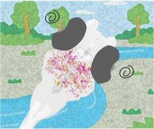
Classic optical illusion leads to the discovery of critical neurons in zebrafish.


By exposing larval zebrafish to a well-known optical illusion, researchers at the Max Planck Institute of Neurobiology and National Institute of Genetics in Japan have found a clever way to isolate key clusters of neurons critical to processing the direction of motion in the zebrafish’s environment.
Details were published in the journal Neuron in September 2020.
Understanding how animal brains are structured and process information is an active area of research that aims to glean insights into fundamental aspects of biological computation. While human brains are seemingly the most impressive animal brain we know of, they are also extremely complex systems containing about as many neurons as there are stars in the Milky Way galaxy, with trillions of connections among them. This complexity makes studying them extremely difficult. Therefore, it is common to study smaller animal brains with much fewer neurons and interconnections, allowing researchers to probe and experiment with simpler- and easier-to-understand brain dynamics.
While smaller animals, such as the larval zebrafish, have simpler and less complex brains, the number of neurons and interconnections is still very large and thus requires researchers to devise experiments that stimulate only small portions of the brain. One way to do this is to stimulate only certain senses, such as sight or taste. This results in specific brain activity, or neural response, to specific inputs.
The research team relied on visual stimuli to study the key neurons necessary for motion processing in the larval zebrafish. By using a well-known optical illusion, the team was able to induce a motion aftereffect (MAE) response in the fish. MAE is found in most animals and arises through viewing a continuous motion in one particular direction, such as a river flowing from the right to left for an extended period of time. If the river is then shut off and the scene becomes still, the animals now sense motion in the reverse direction, from left to right, for a certain period of time following the removal of the flowing river. This indicates the optical illusion that stationary objects in the scene seem to flow in the reverse direction to the motion seen previously.
The team used MAE in the hopes that the perceived motion from the optical illusion would correspond to a smaller set of neural activity that isolates the truly necessary neurons required for motion processing.
Associate Professor Fumi Kubo, a corresponding author of the study, detailed the effectiveness of using MAE to stimulate brain activity in the zebrafish,
“By examining how these numerous neurons respond to an optical illusion, we found that only a dozen or so neurons are required for motion perception in zebrafish larvae.”
Kubo and the team hope to identify how the observed activity fits in with the entire motion processing circuit and study its (inter)connections with the rest of the brain.
Source: Read Full Article