Holliday Junctions in DNA Replication

A holliday junction is a branched nucleic acid structure composed of four double-stranded arms joined together.

kornilov007 | Shutterstock
Holliday junctions can be formed during genetic recombination and affect many biological processes, including DNA replication and gene regulation.
DNA replication
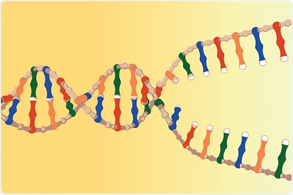
In mitosis, DNA replication is initiated through the actions of various multiprotein complexes that recognize the start sites within a chromosome and cause the separation of DNA into two single-stranded templates.
After this, each DNA strand can be replicated providing two copies of the original DNA. The cell then divides into two to produce two daughter cells containing identical DNA to the parent cell.
DNA replication occurs in three steps:
- Initiation
- Elongation
- Termination
Initiation
Initiation begins at certain points within the DNA known as ‘origins’, and in eukaryotes, this is referred to as the ‘origin recognition complex’ (ORC). ORC is a six-subunit complex, with each subunit interacting with each other and other proteins to form a pre-replication complex which unwinds double-stranded DNA to start replication.
Elongation
The unwinding and replication of DNA creates the replication fork. The replication fork contains both leading and the lagging template strand that are the sites for DNA replication. Elongation is the process which replicates the DNA. DNA polymerases work in a 5’–3’ direction and replicate the DNA on the leading template strand.
The DNA replication on the lagging template strand is performed by discontinuous additions of Okazaki fragments, which involves the placement of a fragment then a short amount of DNA replication followed by the placement of another fragment and so on.
Termination
Eukaryotic DNA replication occurs at many points within the chromosome, therefore replication forks meet and terminate at many points. DNA replication must be stopped or blocked for termination to occur.
To terminate DNA replication, a termination sequence within the DNA and a protein bind to this sequence to stop DNA replication.
Holliday junctions and genetic recombination
Genetic recombination produces daughter cells with combinations of traits that differ from parent cells. During mitosis, the sister chromosomes undergo genetic recombination when near each other, which results in the swapping of alleles between them. This causes a change in the DNA for each daughter cell that is made.
Holliday junctions are a type of DNA cruciform where specific DNA conformations are seen during DNA site-specific recombination, repair, and replication. DNA cruciforms regulate many biological processes that involve DNA, and are formed by inverted nucleotide repeats. They also require DNA supercoiling to remain stable.
Structure of holliday junctions
Holliday junctions take on one of two conformations: folded and unfolded. The unfolded conformation contains adjacent arms that are nearly perpendicular to each other and gives the structure a 4-fold symmetry. The unfolded conformation exists when there is a low concentration of metal ions.
The folded conformation (X-type) has the four arms in a pairwise coaxial stacking structure, which is stabilized by high ionic divalent strength. As the conformation of the holliday junction is dependent on factors, such a metal ion concentration, outside factors have a heavy influence on its conformation.
Purpose of holliday junctions
Holliday junctions regulate gene expression, and they can cause genetic recombination of genes during mitosis. Holliday junctions are targets for many regulatory proteins, such as histones H1 and H5, topoisomerase IIβ, high mobility group (HMG) proteins, and p53.
Many DNA binding proteins, such as HMGB-box family members, BRCA1 protein, and poly ADP ribose polymerase (PARP)-1 show weak binding to DNA but bind very strongly to cruciform structures, such as holliday junctions.
Holliday junctions act as recognition signals near the ORCs of eukaryotic DNA during replication. Other proteins, such as MLL, WRN, 14-3-3, and DEK also show preferential binding.
Sources:
- https://www.ncbi.nlm.nih.gov/pmc/articles/PMC5545932/
- https://www.ncbi.nlm.nih.gov/pmc/articles/PMC5538431/
- https://www.ncbi.nlm.nih.gov/pmc/articles/PMC4404416/
- https://www.ncbi.nlm.nih.gov/pmc/articles/PMC1646289/
- https://www.ncbi.nlm.nih.gov/pmc/articles/PMC3176155/
Further Reading
- All DNA Replication Content
- DNA Replication and Repair
- Homologous Recombination
- Mechanisms of DNA Repair
- Mechanism of DNA Synthesis
Last Updated: Nov 27, 2018
Written by
Samuel Mckenzie
Sam graduated from the University of Manchester with a B.Sc. (Hons) in Biomedical Sciences. He has experience in a wide range of life science topics, including; Biochemistry, Molecular Biology, Anatomy and Physiology, Developmental Biology, Cell Biology, Immunology, Neurology and Genetics.
Source: Read Full Article