Protecting the vision of premature babies


In the spiraling cycle that can lead to vision loss in premature newborns, Medical College of Georgia scientists have found a new target and drug that together appear to stop the destruction in its tracks.
In babies, the development of the blood vessels of the retina should be complete by birth. But with preterm birth, the still-immature retina can develop a potentially blinding eye disorder known as retinopathy of prematurity.
When premature babies transition from inside the womb, where oxygen levels are relatively low, to significantly higher oxygen levels in the incubator, this creates a sensation that their still-developing retina is getting too much oxygen. That sensation inhibits normal blood vessel development, but the retinal neurons keep growing, which leads instead to relative hypoxia, or too little oxygen to the retina.
In the eyes of premature babies as well as people with diabetes, a cascade can follow that should help by growing more blood vessels to make up the oxygen deficit. But the well-intended response can prove problematic instead, resulting in what is called pathological retinal neovascularization.
Now MCG scientists have shown in their animal model of retinopathy of prematurity that the small molecule K604, which is being explored in cancer and Alzheimer’s, can block the development of leaky, obstructive blood vessels in the retina, tamp down inflammation and enable more normal blood vessel growth, ultimately enabling better vision for the babies, they report in the Journal of Neuroinflammation.
K604 blocks ACAT1, or acyl-Coenzyme A: cholesterol acyl transferase 1, an enzyme that converts free cholesterol and long-chain fatty acids to cholesterol esters, basically smaller pieces of cholesterol that can be more easily eliminated by the liver to keep cholesterol levels from getting too high.
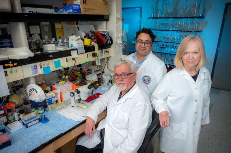
But in premature babies, the hypoxia their retina’s may experience can prompt the formation of dysfunctional blood vessels in the eye and may lead to an accumulation of lipids, fats, and these cholesterol esters, says Modesto A. Rojas, MD, a vascular biologist in the MCG Department of Pharmacology and Toxicology.
It was ACAT1’s role in enabling these toxic cholesterol esters to pile up in the retina that led the MCG scientists to explore what happens when they block it, says Rojas, a corresponding author on the new study.
While cholesterol tends to have a bad reputation, it’s found in most body tissues and is a critical component of the membrane of all cells, including cells in the retina, which capture light and send signals back to the brain, transforming the signals into the images we see. In the altered scenario of the retina of a premature baby, cholesterol starts accumulating and induces inflammation and retinal injury, says first author Syed Adeel H. Zaidi, Ph.D., retina neuroscientist in MCG’s Vascular Biology Center.
Hypoxia increases the expression of the receptor that causes the movement of cholesterol into cells in the retina, which leads to activation of ACAT1 and, consequently, high levels of cholesterol ester. These toxic cholesterol esters promote inflammation, including increased action of TREM1, a receptor in a type of immune cell called myeloid cells that helps turn up short-term inflammation in response to an invader like a virus, Zaidi says.
The scientists have already seen that TREM1 has a role in pathological neovascularization and a relationship with ACAT1. In fact, they have demonstrated TREM1’s inhibition also can essentially prevent problematic neovascularization.
This time they looked at inhibiting ACAT1, which comes before TREM1 in this process, and found it can do the same. Their hypothesis is that ACAT1 inhibition helps restore a more normal metabolism to a situation that has gone haywire.
Part of what goes wrong is that during hypoxia, cholesterol levels increase in the blood as well as the retina, says Zaidi. The scientists reason in the case of their model of retinopathy of prematurity, the microglial cells and resident macrophages make more ACAT1 and cholesterol esters, Rojas says. They suspect the accumulation of cholesterol ester makes these cells more inflammatory.
Microglia are a type of macrophage, immune cells which are adept at ingesting debris and can also help turn inflammation up or down. Microglia are specific to the central nervous system and are always present in the retina to help maintain healthy homeostasis, like keeping the retina clear of debris.
“In hypoxia, microglia/microphages are attracted to the retina by factors released by dying retinal cells,” says Ruth B. Caldwell, Ph.D., cell biologist in the Vascular Biology Center and Culver Vision Discovery Institute at Augusta University, and the study’s senior corresponding author. “They come in and try to digest the resulting debris, but in this hypoxic environment, they don’t just clean; they also get activated.”
“Inflammation is a survival mechanism,” Rojas notes, and microglia are likely trying to help, but their proinflammatory mode just doesn’t get turned off.
As more microglia/macrophages move in, more cholesterol and lipid accumulate, ACAT1 expression increases, and so does the expression of vascular endothelial growth factor, or VEGF, which is needed to make new blood vessels. The unfortunate results include inflammation and growth of abnormal blood vessels, Caldwell says.
The cholesterol keeps coming and ACAT1 keeps metabolizing it because the normal autoregulation to stop making and recruiting cholesterol when levels are sufficient doesn’t work, and the process of producing more cholesterol ester continues, the scientists speculate.
“Cholesterol is taken up more in these proinflammatory microglia/macrophages and when you block the ACAT1 activity, you will see their activation will decrease, so that is how we believe they can still clean things up but be less inflammatory and do more angiogenic repair,” Zaidi says.
In fact, when they removed cholesterol from the equation, it also stopped the destructive cascade, which reiterated cholesterol’s key role in the scenario.
Next steps should include a clinical trial in babies of K604, which is known to be safe in humans, and which, in their model helps dissolve the leaky, obstructive blood vessels paving the way for normal ones, Rojas says.
“This drug is very safe,” he says, and the resulting reduction in abnormal blood vessels comes without altering VEGF levels, he and Caldwell reiterate. Good VEGF levels are needed to enable the growth of sound new blood vessels.
In the lab, the scientists also want to explore the same process they have seen play out in diabetic retinopathy, where high sugars damage previously normal blood vessels in the eye.
Normally excessive cholesterol doesn’t accumulate inside individual cells because ACAT1 keeps converting it to the storage form that can be moved to the liver for elimination from the body. In the retina, at least, ACAT1 doesn’t appear to normally be so busy processing cholesterol, the scientists say.
But another alteration in this dynamic is that expression of the transporters to move the pieces out goes down, so as more cholesterol gets called to the retina it stays there, Zaidi notes.
There is evidence, not unlike with the fat and cholesterol deposited on blood vessel walls with heart disease, that accumulating fat and cholesterol may also play a role in the eye’s misguided efforts to grow new blood vessels, including in patients with diabetic retinopathy where higher levels of the so-called “bad” cholesterol LDL correlate with the activity of macrophages and the severity of the eye disease.
The scientists note that in most premature babies and in many individuals with diabetes, destructive retinopathy does not occur. Diabetic retinopathy occurs in about one-third of patients, and about half of premature babies have some degree of retinopathy of prematurity.
ACAT1 expression and activity in active microglia have been implicated in problems like atherosclerosis, Alzheimer’s and cancer. K604 was developed to work against the excessive cholesterol in atherosclerosis, however it was not effective in that scenario, Caldwell says.
Cholesterol ester levels are currently not measured clinically, like levels of LDL and HDL cholesterol.
More information:
Syed A. H. Zaidi et al, Role of acyl-coenzyme A: cholesterol transferase 1 (ACAT1) in retinal neovascularization, Journal of Neuroinflammation (2023). DOI: 10.1186/s12974-023-02700-5
Journal information:
Journal of Neuroinflammation
Source: Read Full Article