Study assesses T-cell responses directed against highly conserved regions of SARS-CoV-2

Severe acute respiratory syndrome coronavirus 2 (SARS-CoV-2) belongs to the beta-coronavirus family, which is one among the four most common human coronaviruses, beta coronaviruses human coronavirus (HCoV)-OC43 and HCoV-HKU-1, and alpha coronaviruses HCoV-229E and HCoV-NL63.
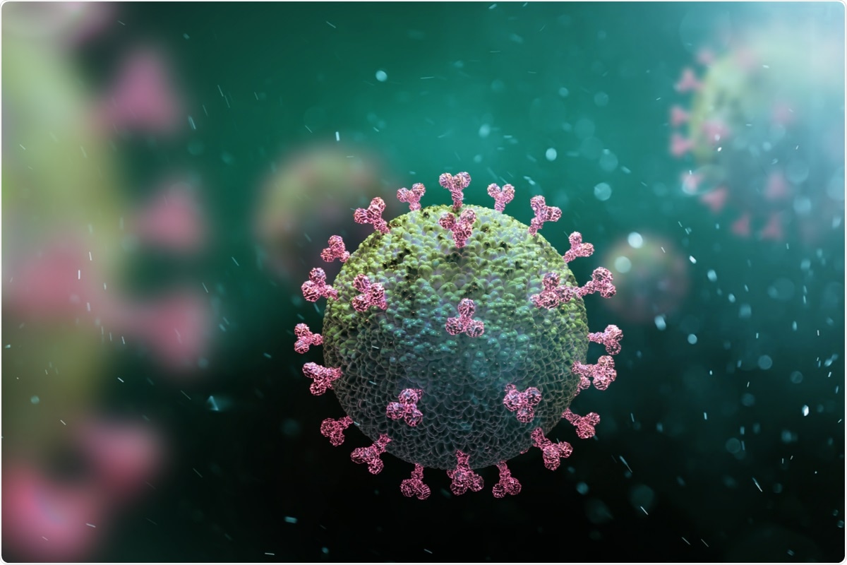
 Study: Low antigen abundance limits efficient T-cell recognition of highly conserved regions of SARS-CoV-2. Image Credit: Andrii Vodolazhskyi/ Shutterstock
Seasonal coronavirus infections typically generate mild symptoms. SARS-CoV-1, SARS-CoV-2, and MERS-CoV infections may have widely variant presentations, ranging from asymptomatic to life-threatening conditions. However, whether a former exposure to common circulating coronavirus provides individuals with immunity against coronavirus disease 2019 (COVID-19) remains debatable.
The genomic structure of SARS-CoV-2 has several open reading frames (ORF) that facilitate virus entry and replication inside the host cells. ORF1ab is a polyprotein involved in viral RNA replication and transcription. Additional ORFs encode the structural proteins, such as nucleocapsid (N), spike (S), envelope (E), and membrane (M), and six accessory proteins ORF3a, ORF6, ORF7a, ORF7b, ORF8, and ORF10. Recent evidence suggests a T-cell immunodominance hierarchy of these antigens.
A preprint version of the study is available on the bioRxiv* server while the article undergoes peer review.
The study
The study aimed to assess whether highly conserved regions within coronaviruses harbor CD8+ T-cell epitopes, capable of being cross-recognized between different coronaviruses, which may provide cross-protection against SARS-COV-2 via pre-existing T-cell immunity.
The study demonstrated that the ORF1ab protein has highly conserved T-cell epitopes that can be recognized by T cells from both COVID-19 convalescent and unexposed participants. However, most human leukocyte antigen (HLA) matched participants may not respond to these epitopes, and the responses are usually subdominant when detectable.
Results
When SARS-Cov2 T-cell epitopes were compared to the corresponding HCoV-OC43 sequences, a maximum of two amino acid changes were depicted.
It was noted that the immunodominant T-cell responses in convalescent individuals primarily targeted ORF3a, N, and S, with limited recognition of ORF1ab-C and nsp12. Most convalescent individuals generated CD8+ T-cell interferon (IFN)-g response above a threshold of 0.5% towards ORF3a, and Spike S1 with 41% reacting to S2.
On the other hand, only 15% of convalescent participants elicited an ORF1ab-C specific CD8+ IFN-g response > 0.5%, while 37% showed an nsp12-specific response. The findings indicated a different pattern of antigen recognition in these donors, with very few showing CD8+ T-cell responses above the 0.5% threshold in response to ORF3a, N, S1, and S2; and more frequent identification of ORF1ab-C and nsp12.
Moreover, a greater proportion of individuals who produced an ORF1ab-C response had mild or no symptoms. On the contrary, participants who did not show a spike protein response had a higher incidence of mild or no symptoms than those with spike responses.
No other potential associations were apparent between the symptoms and response to the other antigens. Additionally, there was no association between the presence of an ORF1ab-C or nsp12 CD8+ T-cell response and the magnitude of the neutralizing antibody response.
Overall, five CD8+ T-cell epitopes were identified, encoded within the ORF1ab-C and nsp12—which included the previously reported SARSCoV-1 and SARS-CoV-2 epitopes FVDGVPFVV (HLA-A*02:07-restricted); LPYPDPSRI (B*51:01- restricted); and DTDFVNEFY (A*01:01-restricted), and two novel epitopes – YAFEHIVY (HLAB35:01-restricted) and NVIPTITQMNL (A*02:05-restricted). In addition, 16 CD8+ immunodominant T-cell epitopes were identified from the ORF3a, N, and S proteins.
T-cell responses were detected against four of the five ORF1ab epitopes in unexposed participants, excluding the HLA-A*01:01 restricted DTD epitope. A majority of B*51:01 positive volunteers responded to the LPY epitope. The T-cell responses to HLA B*35:01-restricted YAF, HLA A*02:05-restricted NVI, and HLA A*02:07-restricted FVD epitopes were rare.
The peptide concentration required to induce 50% maximal activation of CD8+ T cells was calculated and analyzed. The result showed that the immunodominance in these epitopes was not just driven by the selection of T-cell clones with higher functional avidity.
The fine-specificity of CD8+ T cells for their cognate peptide and the complexity of peptide cross-recognition against substitutions at different positions in the peptide sequence was emphasized upon.
However, the nsp10-16 proteins of ORF1ab could not be detected in global proteomic analysis. Nsp10-16 were only detectable following enrichment of RNA-binding proteins, while a high abundance of the ORF3a, N, and S proteins was demonstrated.
Conclusion
In inference, highly conserved regions of coronaviruses provide a source of epitopes for T-cell cross-recognition. Still, limited antigen abundance in infected cells could restrict T-cell priming in coronavirus-exposed individuals. Therefore, widespread protection in the population by pre-existing CD8+ T cells is limited.
*Important notice
bioRxiv publishes preliminary scientific reports that are not peer-reviewed and, therefore, should not be regarded as conclusive, guide clinical practice/health-related behavior, or treated as established information.
- Swaminathan, S. et al. (2021) "Low antigen abundance limits efficient T-cell recognition of highly conserved regions of SARS-CoV-2". bioRxiv. doi: 10.1101/2021.10.13.464181.
Posted in: Medical Science News | Medical Research News | Disease/Infection News
Tags: Amino Acid, Antibody, Antigen, Cell, Coronavirus, Coronavirus Disease COVID-19, Genomic, Human Leukocyte Antigen, immunity, Interferon, Leukocyte, Membrane, MERS-CoV, Protein, Respiratory, RNA, SARS, SARS-CoV-2, Severe Acute Respiratory, Severe Acute Respiratory Syndrome, Spike Protein, Syndrome, T-Cell, Transcription, Virus

Written by
Nidhi Saha
I am a medical content writer and editor. My interests lie in public health awareness and medical communication. I have worked as a clinical dentist and as a consultant research writer in an Indian medical publishing house. It is my constant endeavor is to update knowledge on newer treatment modalities relating to various medical fields. I have also aided in proofreading and publication of manuscripts in accredited medical journals. I like to sketch, read and listen to music in my leisure time.
Source: Read Full Article