Study identifies sex differences in the brain cell types responding to stress


High levels of stress are known to contribute to the development of many psychiatric and medical conditions, including depression, anxiety disorders, hypertension, and heart disease. Past case studies suggest that psychiatric disorders linked to stress exposure can manifest differently in men and women, with the two sexes often predominantly exhibiting different symptoms.
Researchers at the Max Planck Institute of Psychiatry recently carried out a study aimed at better understanding the biological processes underlying these well-documented sex differences in stress responses. Their paper, published in Cell Reports, identifies specific cell types that respond to stress exposure in males and females.
“It is well known that illnesses that arise when people have a high stress load during their life and/or illnesses in which the ability of a person to react to stress is ineffective, show up differently between women and man,” Elena Brivio, one of the researchers who carried out the study, told Medical Xpress.
“For instance, taking depression as an example, we know that almost 2/3 of all depressed patients are women, but we also know that women and man patients develop different sets of symptoms, and they develop them at a different pace (e.g., suicidality and anger is more common in men, apathy and social withdrawal more common in women). However, we do not know the reasons at the molecule and cell level of why these differences exist.”
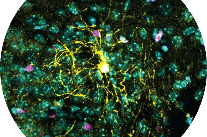
To investigate how the brain of males and females responds to stress, Brivio and her colleagues carried out a series of experiments on mice. In these experiments, they exposed the mice to stress and examined different cell types in a brain region known to coordinate stress responses, namely the paraventricular nucleus within the hypothalamus.
“To study the response to stress of brain cells we used a recent and advanced technique called single-cell RNA sequencing,” Brivio explained. “This technique allows us to quantify which genes—the parts of the DNA that coordinates the life and activity of a cell—are present in the cells. We then checked which genes changed when the animals ware mounting a normal response to stress and if this response was different when the animals were in a chronically stressed condition.”
Using single-cell RNA sequencing, the researchers tried to determine what cells were most sensitive to stress and affected by chronic stress conditions in the brain of male and female mice. They then compared their observations for the two sexes, to unveil similarities or differences between these stress-responsive cell types.
Ultimately, Brivio and her colleagues were able to identify different brain cell types that appear to be involved in male and female stress responses. Their analysis focused on both neurons and glial cells, supporting cells that protect the activity and connections of neurons.
“We found that the most sensitive cell type is a cell called oligodendrocytes, a cell that supports the neurons and allows them to work properly by closely interacting with them,” Brivio said. “Oligodendrocytes showed to be changing if males were exposed to chronic stress, they seemed to look less complex and less mature. But this was true only for males and not females. This tells us that oligodendrocytes might be important cells to be further studied when we try to understand the impact of stress on the brain and its functionality, but we have to keep in consideration that we will likely find something different in males and females.”
This research team gathered new insights about how the brain of men and women reacts to stress, providing a partial biological explanation for previously documented sex differences in the manifestation of disorders linked to stress exposure. While their findings alone are difficult to interpret, they could pave the way for new discoveries, potentially helping to develop more effective sex-specific drugs for stress-induced illnesses.
“In follow-up studies, it would be interesting to better understand how different the oligodendrocytes in the males and females are and the values of the changes we already observed,” Brivio added. “Are the less complex cells we observed in males good news or bad news?
“We could then try to understand which molecular mechanisms lead to these changes and try to prevent them if they are bad news or try to induce them in females if instead are good news. This would allow us to understand if new drugs could be developed targeting these cells and if they could help people suffering from stress-related disorders, or even other disorders.”
The malfunctioning or death of oligodendrocytes has also been linked to other pathologies and medical conditions, thus this recent study could have wider reaching implications. In the future, for instance, it could inform new studies investigating biological sex differences for some of these other conditions, which are still poorly understood.
More information:
Elena Brivio et al, Sex shapes cell-type-specific transcriptional signatures of stress exposure in the mouse hypothalamus, Cell Reports (2023). DOI: 10.1016/j.celrep.2023.112874
Journal information:
Cell Reports
Source: Read Full Article