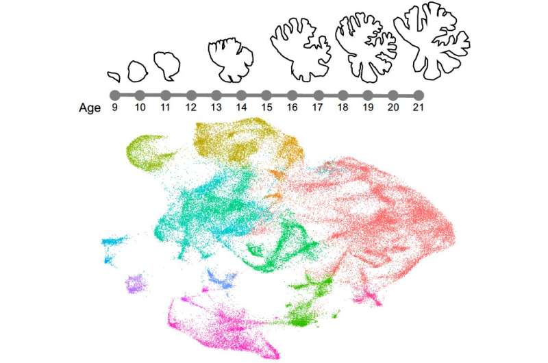
The first molecular map describing human cerebellar development


The cerebellum, a major structure in the human hindbrain, is known to be of central importance for enabling several motor functions, along with cognition, emotional regulation and language processing. Compared to the cerebral cortex, however, this structure is still relatively understudied, and its development over time is poorly understood.
Researchers at Seattle Children’s Research Institute have recently carried out a study aimed at better understanding the development of the human cerebellum. Their paper, published in Nature Neuroscience, offers the first detailed spatial and cell type transcriptional description of the cerebellum’s development.
“We have been in awe of the rich molecular data available to understand the developing human cerebral cortex through resources like BrainSpan and PsychEncode,” Dr Kimberly Aldinger, who carried out the study, told Medical Xpress. “Our goal was to establish a similar resource for the cerebellum for our own studies and to share with the scientific and medical community.”
Dr. Kathleen Millen has spent her research career better understanding cerebellar development. In 2019, research in her lab led by Dr. Parthiv Haldipur and joined by Dr. Aldinger carried out the first comprehensive histological analyses of the developing human cerebellum.
The analyses they conducted allowed them to describe the spatial and temporal expansion of progenitor zones in the human cerebellum and compare these with their development in the rodent brain. Their findings could have significant implications, as they could help to better understand the causes of neurodevelopmental disorders known to affect the cerebellum, such as congenital malformations, autism and pediatric cerebellar cancers.

“I collaborated with the Millen group on the production of this first comprehensive molecular atlas of the cell types and genes expressed during human cerebellar development,” Dr Aldinger said. “This is a rich resource for the scientific community to probe the genetic programs that drive human cerebellar development and origin of disease.”
To conduct their study, Dr Aldinger and her colleagues used SPLiT-seq, a single-cell RNA-sequencing technique developed by researchers at University of Washington (UW) that can be used to assay several cells simultaneously. Using this technique and laser capture technology to achieve the microdissection of spatially defined progenitor regions, the team were able to specifically capture the transcriptome of all developing cerebellar cell types, including very rare cells present in the developing human cerebellum.
“It is hard to model what we don’t know,” Dr Kathleen Millen told Medical Xpress. “Many models of human cerebellar development and disease have been built based on mouse-centric data. Data of the developing human cerebellum was not available, but we now know that human cerebellar development is quite different from that of mice.”
The recent study carried out by this team of researchers led to a number of important findings and achievements. Most notably, the researchers gathered the first molecular data describing the development of the human cerebellum. The data they collected, which is now readily available and can be accessed by other scientists worldwide, could shed some light on the genetic programs enabling normal cerebellar development and the origins of neurodevelopmental disorders associated with abnormal cerebellar development.
The molecular map of human cerebellar development delineated by Dr Aldinger, Dr Millen and their colleagues could soon be used to validate existing mouse and human cerebellar culture models. Remarkably, this map is already allowing both these researchers and other teams to reevaluate their theories about the origins of cerebellar neurodevelopmental disorders.
Source: Read Full Article