Association of self-reported chronic fatigue and retinal microcirculation in post-COVID-19 syndrome patients
In a recent study posted to the medRxiv* preprint server, researchers investigated the potential association between self-reported chronic fatigue (CF) and retinal microcirculation among post-COVID-19 (coronavirus disease 2019) syndrome (PCS) patients.
Background
PCS refers to persistent (>12 weeks) sequelae after severe acute respiratory syndrome coronavirus 2 (SARS-CoV-2) infections, and CF and dyspnoea are the most commonly reported PCS symptoms. Studies have reported that SARS-CoV-2 infection may be caused due to SARS-CoV-2 persistence in blood vessels despite vascular damage, including epitheliopathy, microvasculature impairments, thrombosis, neutrophil extracellular traps (NETs), hypercoagulation, chronic immunological dysregulation, renin-angiotensin-aldosterone-system (RAAS) dysregulations, and autoimmunity/ hyperinflammation as potential PCS pathophysiology mechanisms.
However, PCS pathogenesis has not been well-characterized and requires further investigation. The retinal capillary system is representative of the entire human body’s microcirculation and may represent systemic microvascular changes in systemic diseases. Studies have demonstrated that blood flow impairments could be a potential PCS trigger.
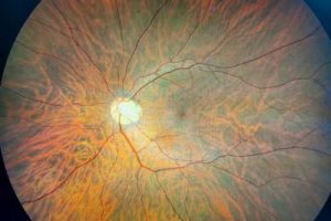
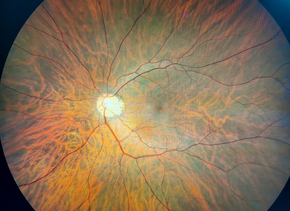
Optical coherence tomography-angiography (OCT-A) enables non-invasive visualization of retinal peripapillary and macular capillary plexi in real time without contacting the human eye, based on temporal intravascular erythrocyte alterations. In addition, the OCT equipment is easy to handle.
About the study
In the present prospective study, researchers investigated if retinal microvasculature circulation could be an objective-type biomarker of the subjectively-reported CF among PCS patients.
The study was conducted at the Ophthalmology department of the University of Erlangen–Nuremberg in Germany and comprised 201 individuals, of which 173 were PCS patients, and 28 were control patients. Retinal microcirculatory assessments were performed based on OCT-A analysis and were quantified as the vascular density (VD) for peripapillary vessels and macula. In addition, participants underwent ophthalmic examinations such as non-contact intraocular pressure (IOP), best-cured visual acuity (BCVA), and axial length measurements.
Self-reported anamnestic CF data were documented, and CF was also assessed based on age, sex, and ‘bell score’ variables. DCP (deep capillary plexus), ICP (intermediate capillary plexus), and SVP (superficial vascular plexus) VD were analyzed 12 times, and mixed modeling was performed to detect potential least square (LS) mean differences between the groups analyzed.
In the first model, PCS patients were compared to controls, with sex and age as covariates. In the second model, the control group was excluded with CF as the independent variable, and the bell score was added as a predictive variable with the same covariates. SARS-CoV-2 infections were confirmed by positive real-time RT-PCR (reverse-transcription-polymerase-chain reaction) analysis reports.
Results and discussion
The average time following SARS-CoV-2 infection was 231 days. Post-COVID symptoms such as CF, concentration difficulties, loss of hair, postural orthostatic tachycardia syndrome (POTS), and subjectively cooler hands were reported by 92%, 83%, 63%, 19%, and 12%, respectively. Among PCS patients, LS means for overall VD were 30, 22, and 24 for SVP, ICP, and DCP, respectively. The corresponding values for controls were 0.1, 22.6, and 23.7, respectively.
Age significantly and inversely impacted SVP, ICP, and DCP VD values between PCS patients and controls, and female PCS patients demonstrated lesser SVP VD than male PCS patients. The first model analysis showed that the ICP VD values were significantly lesser among PCS patients than in the control group. The second model (age, gender, bell scores) analysis revealed significant differences in SVP VD values among PCS patients with CF and those without CF.
PCS patients with PCS and CF showed LS means of VD of 30, 22, and 23 for SVP, ICP, and DCP, respectively. The corresponding VD values for PCS patients without CF were 28, 22, and 24, respectively. SARS-CoV-2 may infect endothelial cells of retinal microvasculature directly via angiotensin-converting enzyme 2 (ACE2) and result in inflammation and fibrosis since ACE2, a serine protease receptor essential for SARS-CoV-2 entry, is situated in the retinal ICP.
Conclusion
Overall, the study findings showed that OCT-A-measured retinal microvascular circulation could be an objective-type biomarker for the subjectively-reported CF among PCS patients, with the human eye, as a diagnostic organ for the entire human body.
*Important notice
medRxiv publishes preliminary scientific reports that are not peer-reviewed and, therefore, should not be regarded as conclusive, guide clinical practice/health-related behavior, or treated as established information.
- Schlick, S. et al. (2022) "Post-COVID-19 syndrome: retinal microcirculation as a potential marker for chronic fatigue". medRxiv. doi: 10.1101/2022.09.23.22280264. https://www.medrxiv.org/content/10.1101/2022.09.23.22280264v1
Posted in: Medical Science News | Medical Research News | Disease/Infection News
Tags: ACE2, Aldosterone, Angiography, Angiotensin, Angiotensin-Converting Enzyme 2, Autoimmunity, Biomarker, Blood, Blood Vessels, Chronic, Coronavirus, Coronavirus Disease COVID-19, covid-19, Diagnostic, Enzyme, Epitheliopathy, Eye, Fatigue, Fibrosis, Hair, Inflammation, OCT, Ophthalmology, Optical Coherence Tomography, Pathophysiology, Polymerase, Receptor, Renin, Respiratory, SARS, SARS-CoV-2, Serine, Severe Acute Respiratory, Severe Acute Respiratory Syndrome, Syndrome, Thrombosis, Tomography, Transcription, Vascular

Written by
Pooja Toshniwal Paharia
Dr. based clinical-radiological diagnosis and management of oral lesions and conditions and associated maxillofacial disorders.
Source: Read Full Article