‘Automated’ microscopy identifies predictor of chemotherapy resistance in ovarian cancer patients


Epithelial ovarian cancer (EOC) is the most common lethal gynecological cancer. Ovarian cancer is usually treated with platinum-based chemotherapy; however, a significant number of patients are resistant to such treatments and relapse soon afterwards. To improve their survival, there is a need to first identify which patients may be platinum-resistant, so that newer treatments may be administered early.
Now, researchers from the Cancer Science Institute of Singapore (CSI Singapore) at the National University of Singapore (NUS), have discovered a way to predict which patients are resistant to platinum chemotherapy. The study, co-led by CSI Singapore Principal Investigators Assistant Professor Anand Jeyasekharan and Associate Professor David Tan, was published in the journal EMBO Molecular Medicine on 11 March 2021.
From their investigation, an association was found between patients whose cancers had high levels of the DNA repair protein, RAD51, and the time to relapse after platinum chemotherapy. “RAD51 has been identified as a biomarker which can potentially be used to determine the resistance of ovarian cancer to platinum chemotherapy,” explained Assoc Prof Tan, who is also a medical oncologist specializing in the treatment of gynecological cancers.
A breakthrough in identifying platinum chemotherapy resistance
RAD51 is a protein that is required for cancers to repair replication-associated DNA damage. Separately, RAD51 is also crucial for repairing platinum chemotherapy-induced damage to the DNA, and the team hypothesized that its overexpression in cancer may therefore affect survival after platinum treatment.
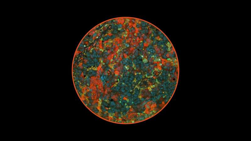
The team used a state‐of‐the‐art automated microscopy method to image and accurately quantify the amount of RAD51 protein present in each tumor cell. Using two independent EOC patient cohorts of 264 and 284 patients from international sites, the researchers showed cases with higher levels of RAD51 relapsed sooner after platinum treatment than those with low levels of RAD51. “This study is the first to use machine-learning based quantitative imaging to measure expression of this DNA repair protein in tumors” said first author Dr. Michal Hoppe, who is a Research Fellow at CSI Singapore.
Importantly, the study also demonstrated that RAD51 overexpression is associated with a unique exclusion of anti-cancer cytotoxic T-cells. “While previous studies have shown associations between loss of DNA repair with changes in the immune microenvironment of cancer, this is the first to our knowledge showing correlation of an increased level of a DNA repair protein with a modified immune response in cancer,” said Asst Prof Jeyasekharan, who is a clinician-scientist.
Next steps
The observation that RAD51 tumors tend to exclude important anti‐cancer immune cells, sets the stage for developing therapeutic approaches to increase immune infiltration in these cancers, and RAD51 expression could then be used to select patients for appropriate treatment.
“The lack of immune cell infiltration into the tumors may explain why these high-RAD51 cancers are more resistant to chemotherapy, and can be further explored as a biomarker to identify patients who may require novel immunotherapy approaches to improve treatment outcomes,” said Assoc Prof Tan.
Furthermore, this study also demonstrates how the use of quantitative molecular imaging can help evaluate the clinical relevance of key changes in the cells that are associated with cancer.
Source: Read Full Article